|
Sphere Fluidics是一家創(chuàng)立于2010年的生命科學公司�����,*初起源于英國劍橋大學��。我們已經(jīng)開發(fā)了40項**產品�����,包括生物芯片和專業(yè)化學制品��,已經(jīng)在全球范圍內超過140名用戶用于研究中�。
我們*初專注于生產新式生物芯片系統(tǒng)和提供研發(fā)服務,現(xiàn)如今我們擴大專家團隊����,正建立一套基于單細胞分析的技術平臺,具備廣闊的市場前景����。我們的系統(tǒng)使得生物藥品的開發(fā)更加快速、更加合算���,改善單克隆抗體的篩選并提高研究效率�����,將為這些工作在診斷和**上帶來激動人心的效益��。
技術簡介:
我們的技術使得篩選百萬個細胞成為可能�,它將為您提供*好的機會來找到稀有的有價值的突變體���,無論它是病變細胞�、有價值的生物產品還是稀有的基因突變體��。我們的技術不僅是**能處理百萬個細胞,而且在高通量篩選和加工應用方面也有**的表現(xiàn)���。
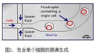
我們**所有的微流控技術使得您能夠超高通量地分析在超小微滴(pL到nL)中的獨立細胞���,這項技術被應用到我們的系統(tǒng)中,從而能夠更加快速���、更低成本并且更有效地進行樣品篩選和生物探索���。
技術優(yōu)勢:
我們的獨特技術讓您能夠在更短的時間內篩選更多的細胞,找到用于**研究的獨一的候選���,生物生產中*佳的克隆和用于研究��、診斷和**的稀有病變細胞���。
通過將微滴技術用于單細胞分析���,您能**處理幾百萬個細胞�。通過將細胞分離到獨立的微滴��,我們的系統(tǒng)保證您更加準確的產物分泌測定。在單克隆抗體方面����,這讓您能夠選擇并分離幾千個抗原特異性的單細胞到獨立的微滴定平板上,同樣的效率在其他生物制品上也是一樣����。
在細胞研究方面,我們理解您的細胞是多么珍貴而脆弱�����,所以我們設計的微滴能在細胞外加一層保護墊��,從而防止細胞受到剪切壓力損傷��。除了技術上的優(yōu)勢�����,我們的系統(tǒng)設計得讓您在實驗室使用簡潔方便��,并且保證清潔無菌����。
產品1:
微滴單細胞包裝系統(tǒng)
簡介:
我們的半自動系統(tǒng)將單細胞或生物分子包裝進微滴中�����,為后續(xù)的篩選和分析做準備����。它的包裝效率可以達到每秒70000個微滴�����,且流速可以嚴格控制�����。這臺設備使得快速且敏感地檢測微滴中單個細胞分泌的或者細胞相關的蛋白成為可能����。
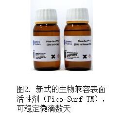
這一包裝系統(tǒng)不影響細胞活性,由于微滴的大小和體積有很大范圍的可變性�,因而能夠用于不同大小的細胞類型。這些微滴被新式的表面活性劑穩(wěn)定�����,細胞能夠在其中生長�,甚至能培養(yǎng)或保存許多天。
主要特點:
·半自動微滴生成器
·在微滴中包裝單個細胞或生物分子
·高速生成微滴(高達70000/秒)
·可同時在微滴中摻入探針和細胞�,使得能夠敏感地檢測到分泌蛋白(例如抗體,生長因子�,細胞因子,酶等)
·用戶決定微流體流動速率
·微滴生成的光學成像經(jīng)過嚴格的質量管理/測試(QA/QC)
·大范圍的微滴大小和體積
應用舉例:
·生物**的發(fā)現(xiàn):從初始漿細胞(B細胞或雜交瘤細胞)中發(fā)現(xiàn)抗體或轉錄產物�����;
·生物加工:快速鑒定和分離高表達克?。?nbsp;
·診斷:探測并測試循環(huán)腫瘤和其他**相關細胞���;
·抗藥性研究:從大量的微生物或腫瘤細胞集群中鑒定和分離稀有的耐藥細胞����;
·酶的進化:篩選數(shù)百萬酶結構以選擇*高效的突變體���;
·合成生物學:研究工程微生物庫中產生的大量有價值的分子���。
技術參數(shù):
規(guī)格
|
|
|
樣品輸入格式
|
注射泵
|
|
樣品輸入體積
|
50μL-1mL
|
|
工作流程
|
生成微滴
|
|
操作環(huán)境
|
|
|
連續(xù)的油相
|
50μL/hr-2000μL/hr
|
|
水相
|
50μL/hr-2000μL/hr
|
|
微滴體積
|
0.2pL-1.7nL
|
|
微滴產率
|
60-70000每秒
|
|
系統(tǒng)規(guī)格
|
|
|
生物芯片兼容性
|
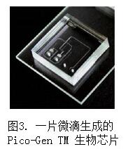
Pico-GenTM微滴生物芯片(更多其他芯片使用請聯(lián)系Sphere Fluidics)
|
|
質量(大約)
|
50kg
|
|
大小(大約)
|
130cmX60cmX60cm(寬X高X深)
|
|
電壓[頻率]
|
110V到240V[@50/60hz]
|
|
能耗
|
300W(*大)
|
|
光學
|
|
|
照明
|
鹵素燈(白光)
|
|
相機
|
高速CMOS(1696X1710像素)
全分辨率下500fps�,弱分辨率下高達200000fps
|
|
相機光譜敏感度
|
400nm-900nm
|
|
PC
|
|
|
電腦
|
Dell Optiplex 7010(4GB RAM;500GB硬盤)或等價物
|
|
PC操作系統(tǒng)
|
Microsoft Windows 7 Professional SP1
|
|
監(jiān)視器
|
彩色LCD(21’’)
|
|
外接
|
2USB��,1以太網(wǎng)
|
|
設備控制軟件
|
neMESYS 注射泵軟件,相機軟件
|
|
工作環(huán)境
|
|
|
間隙
|
30cm
|
|
操作溫度
|
21±5℃
|
產品2:
微滴單細胞測試和分離系統(tǒng)
簡介:
一旦細胞被包裝進微滴中��,這一半自動系統(tǒng)能夠加工��、分選和分離細胞用于簡單分析�。我們知道單細胞分析是多么耗時的,所以我們的系統(tǒng)能夠以高達12000每分鐘的速率加工微滴��,給您更多時間用于分析數(shù)據(jù)�,而不是用于準備準備和加工樣品。
這一系統(tǒng)支持一定范圍的微滴大小和體積�,流速也可控制,同時也配備了可變的光學檢測方法(例如吸光度���、散射光和熒光)����,使之適用于多個研究應用���。
主要特點:
·半自動�、模塊化系統(tǒng)用于分析微滴中的單細胞或生物分子
·高速加工���、檢測和分選微滴(高達12000每分鐘)
·能夠敏感地檢測分泌蛋白(例如抗體�、生長因子、細胞因子和酶等)
·用戶決定微流體流動速率
·微滴生成的光學成像經(jīng)過嚴格的質量管理/測試(QA/QC)
·多種光學檢測方法(例如熒光�、照明和散射)
·大范圍的微滴大小和體積
應用范圍:
·生物**的發(fā)現(xiàn):從初始漿細胞(B細胞或雜交瘤細胞)中發(fā)現(xiàn)抗體或轉錄產物�;
·生物加工:快速鑒定和分離高表達克隆�����;
·診斷:探測并測試循環(huán)腫瘤和其他**相關細胞�����;
·抗藥性研究:從大量的微生物或腫瘤細胞集群中鑒定和分離稀有的耐藥細胞�;
·酶的進化:篩選數(shù)百萬酶結構以選擇*高效的突變體;
·合成生物學:研究工程微生物庫中產生的大量有價值的分子��。
技術參數(shù):
|
規(guī)格
|
|
|
樣品輸入格式
|
注射泵
|
|
樣品輸入體積
|
50μL-1mL
|
|
工作流程
|
生成微滴
|
|
操作環(huán)境
|
|
|
連續(xù)的油相
|
50μL/hr-2000μL/hr
|
|
水相
|
50μL/hr-2000μL/hr
|
|
微滴體積
|
0.2pL-1.7nL
|
|
微滴產率
|
60-70000每秒
|
|
系統(tǒng)規(guī)格
|
|
|
生物芯片兼容性
|
Pico-GenTM微滴生物芯片(更多其他芯片使用請聯(lián)系Sphere Fluidics)
|
|
質量(大約)
|
50kg
|
|
大?。ù蠹s)
|
130cmX60cmX60cm(寬X高X深)
|
|
電壓[頻率]
|
110V到240V[@50/60hz]
|
|
能耗
|
300W(*大)
|
|
光學
|
|
|
照明
|
鹵素燈(白光)
|
|
相機
|
高速CMOS(1696X1710像素)
全分辨率下500fps,弱分辨率下高達200000fps
|
|
相機光譜敏感度
|
400nm-900nm
|
|
PC
|
|
|
電腦
|
Dell Optiplex 7010(4GB RAM�����;500GB硬盤)或等價物
|
|
PC操作系統(tǒng)
|
Microsoft Windows 7 Professional SP1
|
|
監(jiān)視器
|
彩色LCD(21’’)
|
|
外接
|
2USB����,1以太網(wǎng)
|
|
設備控制軟件
|
neMESYS 注射泵軟件�����,相機軟件
|
|
工作環(huán)境
|
|
|
間隙
|
30cm
|
|
操作溫度
|
21±5℃
|
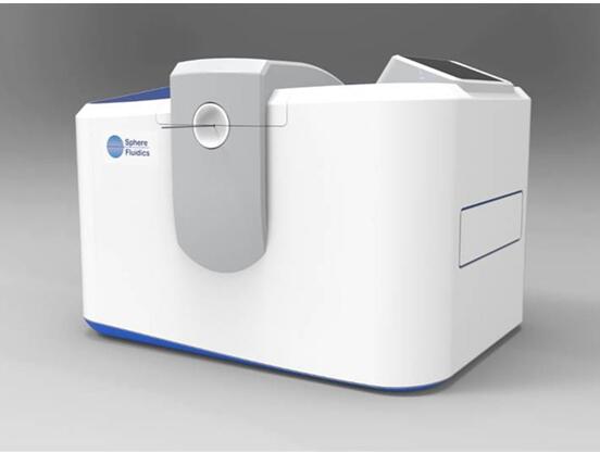
產品3:Cyto-Mine工業(yè)系統(tǒng)(尚未發(fā)布)
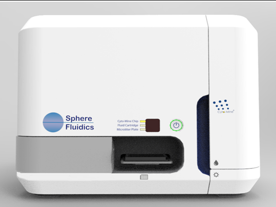
為什么選擇Cyto-Mine �?
很多各色的技術被應用于生物醫(yī)藥的發(fā)現(xiàn)和開發(fā)流程���,包括單細胞分析��、分選����、成像和給微滴定板中的單個孔配藥��。傳統(tǒng)的方式中�����,單個技術需要不同的設備���,這就造成了高的耗費和耗時�,占用了寶貴的實驗室空間��,并且增加了樣品污染的風險。
我們的Cyto-Mine 技術是首款高度整合的設備�����,能夠在單個系統(tǒng)中自動操作所用的重要技術�。這一高通量設備使用微滴和微流體技術,可在**之內加工大約1百萬不同來源的哺乳動物細胞���。每個細胞都被包裹在含有培養(yǎng)基的微滴中,成為區(qū)分細胞并讓其生長的生物反應器�����,*終收獲分泌的分子例如抗體���。這一特殊流程使得選擇性地篩選單個細胞并找到稀有品系成為可能���。
應用范圍:
因為單個細胞是分開的,所以單克隆性是得到保障的����,您可以進行新的測試來測定蛋白分泌率、滴定度和抗原特異性�。Cyto-Mine®中使用的一次性Cyto-Cartridge?保證全程無菌����,減小交叉污染的風險�。這套系統(tǒng)的自動化減少了人力資源的需求,簡單的“載入后運行”模式讓實驗室每個人都能輕松使用�����。
Cyto-Mine®系統(tǒng)有四個主要的應用模式:
1. 單克隆性確保模式:可靠地將單個細胞分選入微滴定板的單孔中�����;
2. 直接測試模式:高通量分選稀有克隆���,并確保*相關的克隆帶上供下游分析的標簽���;
3. 穩(wěn)定性測試模式:及早鑒定不穩(wěn)定克隆,從而在分析中將之去除��,或者在細胞群體中提示您遺傳漂變���;
4. 融合測試模式:分選稀有的細胞-細胞或細胞-生物分子對�。這有利于進行一定的功能測試�,例如B細胞能與分泌特殊抗體的目的細胞共培養(yǎng)���,并造成生成熒光的信號轉導。
產品4:ESI-Mine工業(yè)系統(tǒng)(尚未發(fā)布)
為什么選擇ESI-MineTM�����?
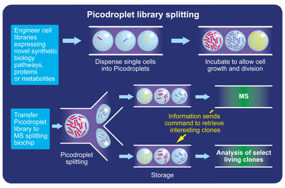
在合成生物學領域中�����,我們的ESI-Mine平臺可以用于高通量質譜(MS)分析����。這一技術能夠檢索在質譜分析中通常被破壞的活拷貝�,當分析稀有蛋白或細胞庫時這點尤其重要。
ESI-Mine?系統(tǒng)首先包裹單個微生物�,然后在可進行代謝和細胞分裂的培養(yǎng)基中培養(yǎng)它們。這些微滴庫隨后轉移到質譜分離生物芯片上����,在那里單個微滴被送往分析。當質譜分析中感興趣的克隆被鑒定出來�,相應的儲存微滴就會被檢索并進行進一步研究。
**的技術:
ESI-Mine?是電噴射電離質譜分析的輔助設備�,每天能夠分析超過200,000個微滴���。這一全自動系統(tǒng)有專用的耗材和軟件,使得其相比于現(xiàn)在的技術運行同樣的樣品測試只用不到10%的時間��。
ESI-Mine技術示意圖
應用實例:
(非參考文獻�,為公司展示資料)
一、初始B細胞的分析
1. 我們的技術如何提高初始B細胞的分析
如果您感興趣的領域與**學相關����,那么您可能經(jīng)歷過分離和篩選B細胞(B**細胞)過程中耗時耗力的體驗。
這一過程可以通過我們的高通量微滴技術得到有效的優(yōu)化����,可以在短時間內篩選大量克隆,增加您在大量細胞集群中找到稀有的有價值的突變體的機會�����。
2. 我們的系統(tǒng)用于初始B細胞分析的獨特優(yōu)勢
將我們的微流體技術用于B細胞分析�,包括動物**和后續(xù)的B細胞分離。有什么重要的優(yōu)勢呢�?通過微滴分選和篩選單個細胞,您能夠自動地分離分泌抗原特異性的抗體進行進一步的分析���。一旦這些細胞被鑒定了����,他們能用于許多應用,包括進一步的測試�����,抗體轉錄產物的RT-PCR���、深度測序和雜交瘤生成�����。
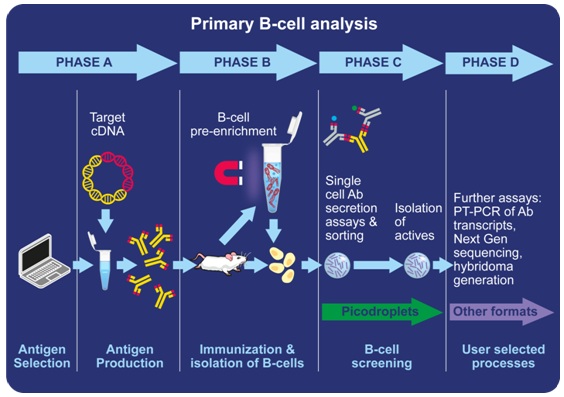
圖1:初始B細胞分析流程圖
3. 我們的系統(tǒng)用于初始B細胞的活動分析
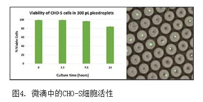
我們都知道整個研究過程中維持細胞活性的重要性��,特別是處理敏感細胞時���。這就是為什么我們的技術需要保證初始B細胞的高活性��,使得細胞在微滴中生長并分析����。如下圖所示,300pL微滴中的細胞在4小時孵育期中保持活性��,大部分的工作流程由我們的系統(tǒng)完成。初始B細胞分析中活性是非常重要的����,在細胞篩選和分選后經(jīng)常也需要保持健康,才能用于下游的應用�。
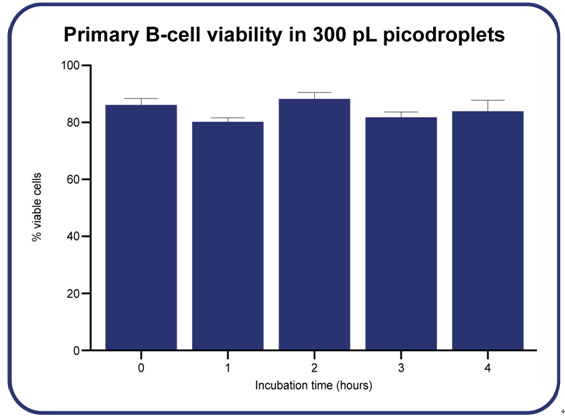
圖2:初始B細胞在300pL微滴中的活性
二、腫瘤細胞**研究
1. 我們的系統(tǒng)如何促進腫瘤研究
腫瘤進展是細胞可造成異常純系擴展的突變導致的結果���,進而導致這些細胞傳播和多樣化����。腫瘤中的細胞通常都不是一樣的�,在大小、蛋白質水平�、基因表達甚至轉移的能力都各有不同,這就導致我們很難理解是哪些細胞導致腫瘤的生長和多樣化��,以及是哪些因素影響它們與周邊環(huán)境作用���,這些問題*大地困擾了相關研究者們�����。
在Sphere�,我們考慮到這些困難,并發(fā)展出一個獨特的平臺��,幫助您篩選單個細胞�����,因而您能夠研究單個細胞的基因和蛋白質表達譜��,找到新的調控通路或者先前未檢測到的克隆并進一步研究���。
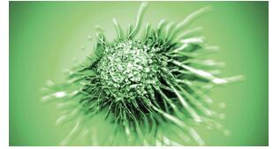
圖1:腫瘤細胞圖示
2. 我們的系統(tǒng)用于腫瘤研究的獨特優(yōu)勢
循環(huán)的腫瘤細胞的比例大概是一個對一百萬個白細胞���,或者一個對十億個紅細胞。傳統(tǒng)的實驗中�,從復雜來源的細胞群中分離出腫瘤細胞非常耗時,并且技術上非常困難�����。
幸運的是�,我們的實驗儀器能夠高通量地分離單細胞�。我們新穎的技術使用微滴包裹從臨床樣本中分離出來的白細胞,并進行高通量分析����。這些分離出來的單個腫瘤細胞能夠被進一步地鑒定和分析����,從而為癌癥患者開發(fā)并精煉個性化的**���。我們的技術也有助于單細胞蛋白質組的分析�,以及下游的基因組和表觀基因組分析��,進而解析液態(tài)活組織的腫瘤細胞群���。
圖2:我們的系統(tǒng)圖示
重要圖解:
英文介紹:
Our research instruments are designed to assist you in finding highly valuable and rare biological variants among vast cell populations. By increasing speed and reducing cost, our picodroplet technology can help you save resources whilst boosting your chances of success.
Unlike our industrial instruments, our research instruments are semi-automated rather than fully automated. While this means they require slightly more user input to configure, it also means that they are more flexible and can be easily adapted to fit the needs of your unique research project.
We currently have two research instruments that enable you to generate, isolate, and dispense picodroplets for a range of applications. These are the Picodroplet Single Cell Encapsulation System and the Picodroplet Single Cell Assay and Isolation System. Both are compatible with our range of specialist chemicals and biochips.
Some common research areas where our instruments have been used previously include biopharmaceutical discovery, bioprocessing, diagnostics, drug-resistance studies, enzyme evolution and synthetic biology.
Scroll down for more information on our instruments, or alternatively, contact one of our experts to discuss your needs.
Picodroplet Single Cell Encapsulation System
Our semi-automated system encapsulates single cells or biomolecules into picodroplets, ready for downstream screening and analysis. It does this at a rapid rate of up to 70,000 picodroplets per second, and the flow rate can be highly controlled. The instrument enables rapid and very sensitive detection of secreted or cell-associated proteins produced by an individual cell, contained in each picodroplet.
The encapsulation system doesn’t affect cell viability, while the wide range of picodroplet sizes and volumes gives it flexibility for use with various cell types – large or small. These picodroplets can be stabilised using novel surfactants and cells can be grown in them, and even incubated or stored for many days.
Download the Picodroplet Single Cell Encapsulation System information leaflet to learn more:
Download Leaflet to Learn More
Picodroplet Single Cell Assay and Isolation System
Once the cells have been encapsulated in picodroplets, this semi-automated system is able to process, sort and isolate them for easy analysis. We know how time-consuming single cell analysis can be, and that’s why our system is capable of processing picodroplets at a rate of up to 12,000 per minute – giving you more time to spend analysing your data rather than on preparing and processing your samples.
The system supports a range of picodroplet sizes and volumes, and the flow rate can be controlled. It’s also equipped with various optical detection methods (e.g. absorbance, scatter, fluorescence) to make it suitable for a range of research applications.
Download the Picodroplet Single Cell Assay and Isolation System information leaflet to learn more:
應用文獻集
1. Abalde-Cela et al.
2015. High-throughput detection of ethanol-producing cyanobacteria in a
microdroplet platform. J. R. Soc. Interface 12: 2015.0216
2. Bakewell
et al. 2015. Information processing tools for extracting the electrical
properties of nanoparticles. AIP Conf. Proc. 1646, 17-24
3. Bakewell
et al. Exploring and Evaluating Micro-environment and Nanoparticle
Dielectrophoretic-induced Interactions with Image Analysis Methods. Materials
Today: Proceedings, 867-874, 3(3) 2016.
4. Chokkalingam
et al. 2013. Probing cellular heterogeneity in cytokine-secreting immune cells
using droplet-based microfluidics. Lab Chip 13: 4740-4744
5. Holmes et
al. 2014. Separation of blood cells with differing deformability using
deterministic lateral displacement. Interface Focus 4. 20140011
6. Kruger
et al. 2014. Deformability-based red blood cell separation in deterministic
lateral displacement devices—A simulation study. Biomicrofluidics 8; 054114
7. Ma et
al. 2012. Fabrication of Microgel Particles with Complex Shape via Selective
Polymerization of Aqueous Two-Phase Systems. Small. 8(15): 2356-2360
8. Ma et al. 2013. Monodisperse collagen–gelatin
beads as potential platforms for 3D cell culturing. J. Mater. Chem. B, 1;
5128-5136
9.Salmon
et al. 2016. Monitoring early-stage nanoparticle assembly in microdroplets by
optical spectroscopy and SERS. Small. Doi:10.1002/smll.201503513
10.
Sherwood et al. 2014. Spatial Distributions of Red Blood Cells Significantly
Alter Local Haemodynamics. PLOS One 9(6): :e100473
11. Shim et al. 2013.
Ultrarapid Generation of Femtoliter Microfluidic Droplets for
Single-Molecule-Counting Immunoassays. ACS Nano 7(7): 5955-5964
12. Smith et al. 2013.
Sensitive, High Throughput Detection of Proteins in Individual,
Surfactant-Stabilized Picoliter Droplets Using Nanoelectrospray Ionization Mass
Spectrometry. Analytical Chemistry. 85(8): 3812-3816
13. Parker, R. M. et
al. 2015. Electrostatically directed self-assembly of ultrathin supramolecular
polymer microcapsules. Advanced Functional Materials. 25(26): 4091-4100.
14. Use
of standards for digital biological information in the design, construction and
description of a synthetic biological system – Guide. 2015. PAS 246:2015
|