|
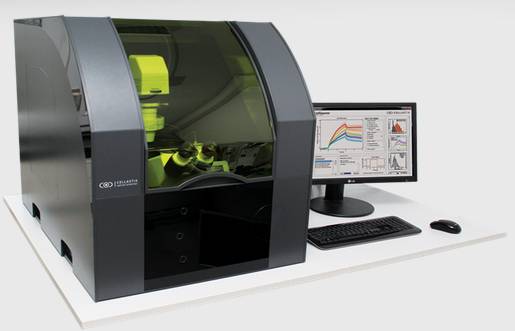
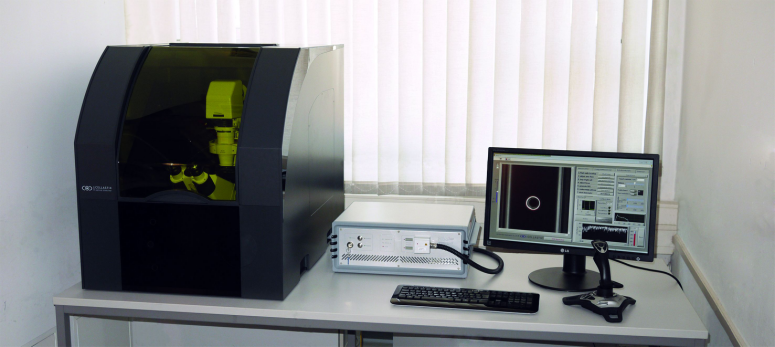
單細胞牽張拉伸壓縮形變與機械力特性測試分析系統(tǒng)
——基于微流控技術(shù)的單細胞機械力特性精準(zhǔn)��、高通量表征
Single cell mechanics - the easy way
背景:
單細胞水平的機械力特性表征��,可以有效闡明細胞的功能和狀態(tài)��,揭示細胞的單體差異性����,對于細胞的分化和病理研究���,以及**的早期臨床診斷和**具有非常重要的意義。 該系統(tǒng)基于微流控芯片的方式更適合單細胞樣本的微環(huán)境**控制��、高通量定向操縱及多參數(shù)非特異性檢測�。
簡介:
該單細胞高通量牽張與力學(xué)特性測試分析系統(tǒng),是****臺用來高通量測量、分析單個懸浮細胞形變的設(shè)備�����。用來可視化研究討論細胞力學(xué)性質(zhì)與其功能之間的關(guān)系 該系統(tǒng)可以安裝在任何相位差顯微鏡上的模塊����。溫度穩(wěn)定和激光**。

  
系統(tǒng)亮點特性:
1 )可大量表征單細胞機械力特性����、操作簡便、樣品消耗量小
該系統(tǒng)的微流控芯片具有與細胞直徑良好相符性的微納米級腔道����,并能實現(xiàn)對微流體的**控制,使其尤其適合單細胞機械特性研究分析�����,該微流控的高通量技術(shù)便于大量表征單細胞機械力特性、操作簡便����、樣品消耗量小、集成和微型化程度高等優(yōu)點�,且在分析過程中單細胞懸浮高速流經(jīng)檢測區(qū)域,該連續(xù)流動態(tài)檢測的特性*大提高了系統(tǒng)的通量���。
2)高速對單個細胞進行形變�����,并進行機械特性高速表征��,單細胞高通量流變
利用兩素未聚焦光進行單細胞形變��,并通過圖形化微柱基地表征細胞的力特性,高速有效分析單細胞水平的機械特性, 高達300個細胞/小時.
3)非機械接觸�����、無標(biāo)記進行細胞捕捉和拉伸,確保細胞**與細胞損傷*小化(Contact-free cell deformation)
利用光延伸器技術(shù)測試細胞機械特性能時���,在非機械接觸情況下細胞進行捕捉和拉伸���,且不需要對激光進行聚焦�,能實現(xiàn)細胞損傷*小化����。優(yōu)于AFM(原子力顯微鏡)和光鑷
4)將光延伸器**性與微流控高通量**相結(jié)合,細胞機械特性測試分析**而且高效
采用2個微流道來輸送細胞����,使兩條光纖垂直分布于通道兩側(cè)并嚴格對準(zhǔn)? ,單細胞隨流體進入檢測區(qū)域時����,首先采用功率較低的光速捕獲細胞,然后增加光速的功率使細胞發(fā)生形變�����。通過對細胞變形能力的分析�,不僅能區(qū)分病變細胞和正常細胞,而且可以用于辨別轉(zhuǎn)型特性和非轉(zhuǎn)移特性的癌細胞��。
5)自動化測量單細胞力屬性和成像記錄細胞形變記錄
對應(yīng)于用戶定義的拉伸模式�,細胞被自動傳送到測量區(qū)域由CellStretcher模塊控制所有組件和自動測量細胞;細胞形變由系統(tǒng)CCD相機自動記錄��,并由CellEvaluator自動提取記錄顯微圖像形變數(shù)據(jù),CellReporter可視化統(tǒng)計分析表征參數(shù)�。在光學(xué)拉伸加載運行實驗中,科研學(xué)者可專注于闡述實驗結(jié)果
6)良好溫控微環(huán)境罩


Publications
RS ZELLTECHNIK BROCHURES
The Optical Stretcher
OPTICAL STRETCHER TECHNOLOGY
Lincoln, B., Schinkinger, S., Travis, K., Wottawah, F., Ebert, S., Sauer, F., Guck, J., 2007. Reconfigurable microfluidic integration of a dual-beam laser trap with biomedical applications. Biomed. Microdevices 9, 703–710. doi:10.1007/s10544-007-9079-x
Ebert, S., Travis, K., Lincoln, B., Guck, J., 2007. Fluorescence ratio thermometry in a microfluidic dual-beam laser trap. Opt. Express 15, 15493–15499. doi:10.1364/OE.15.015493
Jensen-McMullin, C., Lee, H.P., Lyons, E.R.L., 2005. Demonstration of trapping, motion control, sensing and fluorescence detection of polystyrene beads in a multi-fiber optical trap. Opt. Express 13, 2634–2642. doi:10.1364/OPEX.13.002634
Wottawah, F., Schinkinger, S., Lincoln, B., Ananthakrishnan, R., Romeyke, M., Guck, J., K?s, J., 2005. Optical Rheology of Biological Cells. Phys. Rev. Lett. 94, 098103. doi:10.1103/PhysRevLett.94.098103
Lincoln, B., Erickson, H.M., Schinkinger, S., Wottawah, F., Mitchell, D., Ulvick, S., Bilby, C., Guck, J., 2004. Deformability-based flow cytometry.Cytometry A 59A, 203–209. doi:10.1002/cyto.a.20050
THEORETICAL MODELS
Ananthakrishnan, R., Guck, J., Wottawah, F., Schinkinger, S., Lincoln, B., Romeyke, M., Kas, J., 2005. Modelling the structural response of an eukaryotic cell in the optical stretcher. Curr. Sci. 88.
B. Bareil, P., Sheng, Y., Chiou, A., 2006. Local scattering stress distribution on surface of a spherical cell in optical stretcher. Opt. Express 14, 12503–12509. doi:10.1364/OE.14.012503
Bareil, P.B., Sheng, Y., Chen, Y.-Q., Chiou, A., 2007. Calculation of spherical red blood cell deformation in a dual-beam optical stretcher. Opt. Express 15, 16029–16034. doi:10.1364/OE.15.016029
Boyde, L., Ekpenyong, A., Whyte, G., Guck, J., 2012. Comparison of stresses on homogeneous spheroids in the optical stretcher computed with geometrical optics and generalized Lorenz–Mie theory. Appl. Opt. 51, 7934–7944. doi:10.1364/AO.51.007934
Ekpenyong, A.E., Posey, C.L., Chaput, J.L., Burkart, A.K., Marquardt, M.M., Smith, T.J., Nichols, M.G., 2009. Determination of cell elasticity through hybrid ray optics and continuum mechanics modeling of cell deformation in the optical stretcher. Appl. Opt. 48, 6344–6354. doi:10.1364/AO.48.006344
Teo, S.-K., Goryachev, A.B., Parker, K.H., Chiam, K.-H., 2010. Cellular deformation and intracellular stress propagation during optical stretching.Phys. Rev. E 81, 051924. doi:10.1103/PhysRevE.81.051924
CANCER RESEARCH AND DIAGNOSTICS
Kastl, L., Budde, B., Isbach, M., Rommel, C., Kemper, B., Schnekenburger, J., 2015. Optomechanical properties of cancer cells revealed by light-induced deformation and quantitative phase microscopy. pp. 952908–952908–6. doi:10.1117/12.2184764
Martin, M., Müller, K., Cadenas, C., Hermes, M., Zink, M., Hengstler, J.G., K?s, J.A., 2012. ERBB2 overexpression triggers transient high mechanoactivity of breast tumor cells. Cytoskeleton 69, 267–277. doi:10.1002/cm.21023
Fritsch, A., H?ckel, M., Kiessling, T., Nnetu, K.D., Wetzel, F., Zink, M., K?s, J.A., 2010. Are biomechanical changes necessary for tumour progression? Nat. Phys. 6, 730–732. doi:10.1038/nphys1800
Brunner, C., Niendorf, A., K?s, J.A., 2009. Passive and active single-cell biomechanics: a new perspective in cancer diagnosis. Soft Matter 5, 2171–2178. doi:10.1039/B807545J
Remmerbach, T.W., Wottawah, F., Dietrich, J., Lincoln, B., Wittekind, C., Guck, J., 2009. Oral Cancer Diagnosis by Mechanical Phenotyping. Cancer Res. 69, 1728–1732. doi:10.1158/0008-5472.CAN-08-4073
Martin, M., Mueller, K., Wottawah, F., Schinkinger, S., Lincoln, B., Romeyke, M., K?s, J.A., 2006. Feeling with light for cancer. p. 60800P–60800P–10. doi:10.1117/12.637899
Guck, J., Schinkinger, S., Lincoln, B., Wottawah, F., Ebert, S., Romeyke, M., Lenz, D., Erickson, H.M., Ananthakrishnan, R., Mitchell, D., K?s, J., Ulvick, S., Bilby, C., 2005. Optical Deformability as an Inherent Cell Marker for Testing Malignant Transformation and Metastatic Competence. Biophys. J. 88, 3689–3698. doi:10.1529/biophysj.104.045476
STEM CELL RESEARCH
Ekpenyong, A.E., Whyte, G., Chalut, K., Pagliara, S., Lautenschlaeger, F., Fiddler, C., Paschke, S., Keyser, U.F., Chilvers, E.R., Guck, J., 2012.Viscoelastic Properties of Differentiating Blood Cells Are Fate- and Function-Dependent. Plos One 7, e45237. doi:10.1371/journal.pone.0045237
Galle, J., Bader, A., Hepp, P., Grill, W., Fuchs, B., Kas, J.A., Krinner, A., MarquaB, B., Muller, K., Schiller, J., Schulz, R.M., von Buttlar, M., von der Burg, E., Zscharnack, M., Loffler, M., 2010. Mesenchymal Stem Cells in Cartilage Repair: State of the Art and Methods to monitor Cell Growth, Differentiation and Cartilage Regeneration. Curr. Med. Chem. 17, 2274–2291. doi:10.2174/092986710791331095
Maloney, J.M., Nikova, D., Lautenschlager, F., Clarke, E., Langer, R., Guck, J., Van Vliet, K.J., 2010. Mesenchymal Stem Cell Mechanics from the Attached to the Suspended State. Biophys. J. 99, 2479–2487. doi:10.1016/j.bpj.2010.08.052
Lautenschl?ger, F., Paschke, S., Schinkinger, S., Bruel, A., Beil, M., Guck, J., 2009. The regulatory role of cell mechanics for migration of differentiating myeloid cells. Proc. Natl. Acad. Sci. 106, 15696–15701 doi:10.1073/pnas.0811261106
IMMUNE SYSTEM
Man, S.M., Ekpenyong, A., Tourlomousis, P., Achouri, S., Cammarota, E., Hughes, K., Rizzo, A., Ng, G., Wright, J.A., Cicuta, P., Guck, J.R., Bryant, C.E., 2014. Actin polymerization as a key innate immune effector mechanism to control Salmonella infection. Proc. Natl. Acad. Sci. 201419925 doi:10.1073/pnas.1419925111
BASIC RESEARCH
Schmidt, B.U.S., Kie?ling, T.R., Warmt, E., Fritsch, A.W., Stange, R., K?s, J.A., 2015. Complex thermorheology of living cells. New J. Phys. 17, 073010. doi:10.1088/1367-2630/17/7/073010
Chan, C.J., Ekpenyong, A.E., Golfier, S., Li, W., Chalut, K.J., Otto, O., Elgeti, J., Guck, J., Lautenschl?ger, F., 2015. Myosin II Activity Softens Cells in Suspension. Biophys. J. 108, 1856–1869. doi:10.1016/j.bpj.2015.03.009
Gladilin, E., Gonzalez, P., Eils, R., 2014. Dissecting the contribution of actin and vimentin intermediate filaments to mechanical phenotype of suspended cells using high-throughput deformability measurements and computational modeling. J. Biomech. 47, 2598–2605. doi:10.1016/j.jbiomech.2014.05.020
Maloney, J.M., Vliet, K.J.V., 2014. Chemoenvironmental modulators of fluidity in the suspended biological cell. Soft Matter. doi:10.1039/C4SM00743C
Warmt, E., Kie?ling, T.R., Stange, R., Fritsch, A.W., Zink, M., K?s, J.A., 2014. Thermal instability of cell nuclei. New J. Phys. 16, 073009. doi:10.1088/1367-2630/16/7/073009
Gyger, M., Stange, R., Kiessling, T.R., Fritsch, A., Kostelnik, K.B., Beck-Sickinger, A.G., Zink, M., Kaes, J.A., 2014. Active contractions in single suspended epithelial cells. Eur. Biophys. J. Biophys. Lett. 43, 11–23. doi:10.1007/s00249-013-0935-8
Seltmann, K., Fritsch, A.W., K?s, J.A., Magin, T.M., 2013. Keratins significantly contribute to cell stiffness and impact invasive behavior. Proc. Natl. Acad. Sci. 201310493. doi:10.1073/pnas.1310493110
Maloney, J.M., Lehnhardt, E., Long, A.F., Van Vliet, K.J., 2013. Mechanical fluidity of fully suspended biological cells. Biophys. J. 105, 1767–1777. doi:10.1016/j.bpj.2013.08.040
Kie?ling, T.R., Stange, R., K?s, J.A., Fritsch, A.W., 2013. Thermorheology of living cells—impact of temperature variations on cell mechanics. New J. Phys. 15, 045026. doi:10.1088/1367-2630/15/4/045026
Kie?ling, T.R., Herrera, M., Nnetu, K.D., Balzer, E.M., Girvan, M., Fritsch, A.W., Martin, S.S., K?s, J.A., Losert, W., 2013. Analysis of multiple physical parameters for mechanical phenotyping of living cells. Eur. Biophys. J. 42, 383–394. doi:10.1007/s00249-013-0888-y
Paschke, S., Weidner, A.F., Paust, T., Marti, O., Beil, M., Ben-Chetrit, E., 2013. Technical advance: Inhibition of neutrophil chemotaxis by colchicine is modulated through viscoelastic properties of subcellular compartments. J. Leukoc. Biol. 94, 1091–1096. doi:10.1189/jlb.1012510
Chalut, K.J., H?pfler, M., Lautenschl?ger, F., Boyde, L., Chan, C.J., Ekpenyong, A., Martinez-Arias, A., Guck, J., 2012. Chromatin decondensation and nuclear softening accompany Nanog downregulation in embryonic stem cells. Biophys. J. 103, 2060–2070. doi:10.1016/j.bpj.2012.10.015
Matthews, H.K., Delabre, U., Rohn, J.L., Guck, J., Kunda, P., Baum, B., 2012. Changes in Ect2 localization couple actomyosin-dependent cell shape changes to mitotic progression. Dev. Cell 23, 371–383. doi:10.1016/j.devcel.2012.06.003
Mauritz, J.M.A., Esposito, A., Tiffert, T., Skepper, J.N., Warley, A., Yoon, Y.-Z., Cicuta, P., Lew, V.L., Guck, J.R., Kaminski, C.F., 2010. Biophotonic techniques for the study of malaria-infected red blood cells. Med. Biol. Eng. Comput. 48, 1055–1063. doi:10.1007/s11517-010-0668-0
Rusciano, G., 2010. Experimental analysis of Hb oxy–deoxy transition in single optically stretched red blood cells. Phys. Med. 26, 233–239. doi:10.1016/j.ejmp.2010.02.001
AGING PROCESSES
Schulze, C., Wetzel, F., Kueper, T., Malsen, A., Muhr, G., Jaspers, S., Blatt, T., Wittern, K.-P., Wenck, H., K?s, J.A., 2010. Stiffening of Human Skin Fibroblasts with Age. Biophys. J. 99, 2434–2442. doi:10.1016/j.bpj.2010.08.026
VESICLES
Delabre, U., Feld, K., Crespo, E., Whyte, G., Sykes, C., Seifert, U., Guck, J., 2015. Deformation of phospholipid vesicles in an optical stretcher. Soft Matter. doi:10.1039/C5SM00562K
Solmaz, M.E., Sankhagowit, S., Biswas, R., Mejia, C.A., Povinelli, M.L., Malmstadt, N., 2013. Optical stretching as a tool to investigate the mechanical properties of lipid bilayers. Rsc Adv. 3, 16632–16638. doi:10.1039/c3ra42510j
Solmaz, M.E., Biswas, R., Sankhagowit, S., Thompson, J.R., Mejia, C.A., Malmstadt, N., Povinelli, M.L., 2012. Optical stretching of giant unilamellar vesicles with an integrated dual-beam optical trap. Biomed. Opt. Express 3, 2419–2427. doi:10.1364/BOE.3.002419
TECHNICAL ADVANCES
Grosser, S., Fritsch, A.W., Kie?ling, T.R., Stange, R., K?s, J.A., 2015. The lensing effect of trapped particles in a dual-beam optical trap. Opt. Express 23, 5221–5235. doi:10.1364/OE.23.005221
Bellini, N., Bragheri, F., Cristiani, I., Guck, J., Osellame, R., Whyte, G., 2012. Validation and perspectives of a femtosecond laser fabricated monolithic optical stretcher. Biomed. Opt. Express 3, 2658–2668. doi:10.1364/BOE.3.002658
Bellini, N., Vishnubhatla, K.C., Bragheri, F., Ferrara, L., Minzioni, P., Ramponi, R., Cristiani, I., Osellame, R., 2010. Femtosecond laser fabricated monolithic chip for optical trapping and stretching of single cells. Opt. Express 18, 4679–4688. doi:10.1364/OE.18.004679
|